. 2017 Dec 11;9(5):1279–1288. doi: 10.1039/c7sc04521b

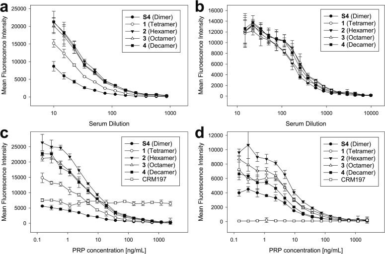
Fig. 4. Glycan array binding analysis of anti-PRP antibodies to sPRP oligosaccharides 1–4. (a and b) Microarray slides printed with sPRP were incubated with a dilution series of a human reference serum (a) and rabbit anti-Hib antiserum (b) followed by a fluorescent secondary antibody, and fluorescence intensities for the synthetic length-defined glycans were determined. (c and d) To verify specificity of the analyzed antibodies, antibody binding was inhibited competitively by adding different concentrations of native PRP to human reference serum diluted 1 : 200 (c) or rabbit typing serum diluted 1 : 1000 (d). All values are the mean of eight spots from two replicate wells with error bars denoting the standard deviation (SD).

Fig. 4