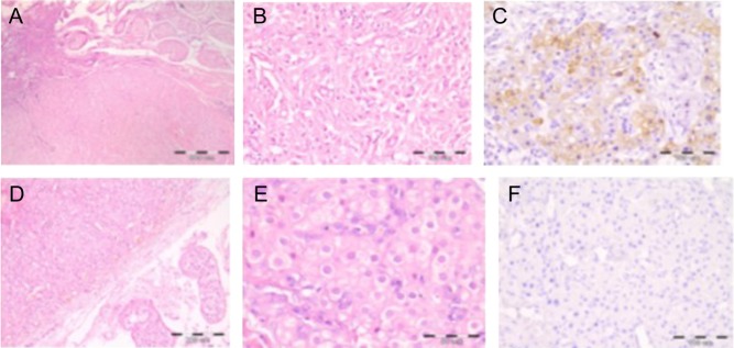
Figure 4.
Histology of the testes in the two biopsied patients. Upper line: Patient 1. (A) The arrow points to the delineation between adrenal and testicular tissue. (B) Larger view. (C) Positive inhibin staining. Lower line: Patient 3. (D) Adrenal tissue in the testis (magnification ×400). (E) Closer image of the TART tissue. (F) Negative inhibin staining.

 This work is licensed under a