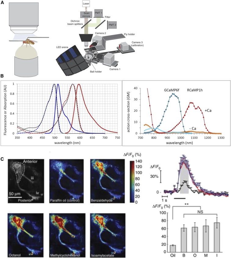
Figure 1.
Functional imaging in the adult fly brain with GCaMP. (A) Schematic of a two-photon imaging rig, showing a tethered fly walking on a ball with a window cut in the head for visualization of GCaMP fluorescence changes in the brain. This diagram is taken from figure 1 in Seelig et al. (2010); alternative schematics are shown in Reiff et al. (2010), Maimon et al. (2010), and Mamiya and Dickinson (2015). (B) Excitation and emission spectra of GCaMP and RCaMP (Chen et al. 2013; Dana et al. 2016), taken from https://www.janelia.org/lab/harris-lab-apig/research/photophysics/two-photon-fluorescent-probes. (C) Examples of ways to show changes in GCaMP fluorescence in mushroom body neurons in response to odors [taken from figure 6 in Sejourne et al. (2011)]. AU, arbitrary units; GM, Goeppert-Mayer Units; LED, light-emitting diode; **, very significant P value between 0.001 and 0.01; NS, not significant; PMT, photomultiplier tube detector.

