
Fig. 3.
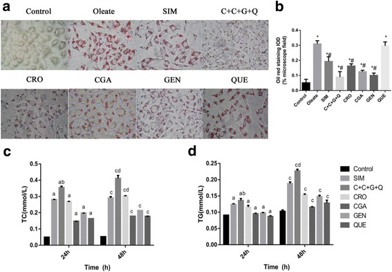
The effects of CRO, CGA, GEN, QUE and C + C + G + Q on lipid content in HepG2 cells. HepG2 cells were treated with Oleate (0.1 mmol/L), SIM (10 μmol/L), CRO (1 μmol/L), CGA (30 μmol/L), GEN (10 μmol/L), QUE (10 μmol/L) and C + C + G + Q (1 μmol/L + 30 μmol/L + 10 μmol/L + 10 μmol/L) for 24 h or 48 h. a After 48 h incubation with different drugs, the images of cells were observed by microscope at 250 × original magnification showing lipid accumulation in cells stained by Oil red O. b The comparison of integral optical density (IOD) for Oil red staining in cells. Effects of different drugs on the secretion of (c) TC and (d) TG were observed by HepG2 cells in minimum essential medium. Data are expressed as mean ± SEM from at least four independent experiments. *P < 0.05 vs. control, #P < 0.05 vs. Oleate. After 24 h, aP < 0.05 vs. control, bP < 0.05 vs. SIM. After 48 h, cP < 0.05 vs. control, dP < 0.05 vs. SIM