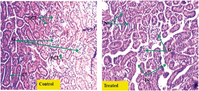
Fig. 5.
Histopathological changes in kidney tissue after Cr(VI) exposure. Representative hematoxylin and eosin-stained histological sections from kidney of vehicle-treated normal renal cortex (Control) (magnification 20×; G, glomerulus; PCT, proximal convoluted tubule; DCT, distal convoluted tubule; CT, collecting tubule); and (Treated) chromium-treated mice renal cortex [magnification 20×: tubular damage with mild tubular dilatation, necrosis of tubular epithelial cells (N) and increased interstitial spaces (IS)].

