Figure 2.

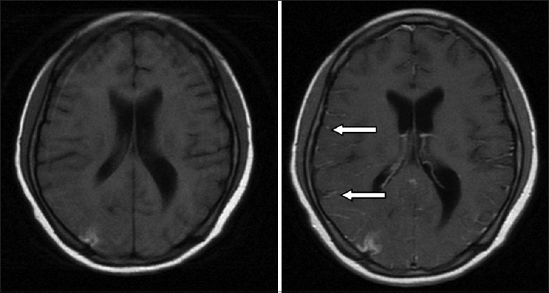
The left picture is from normal magnetic resonance imaging and the right picture is from enhanced magnetic resonance imaging. The leptomeninges are enhanced on the right picture (white arrow).

The left picture is from normal magnetic resonance imaging and the right picture is from enhanced magnetic resonance imaging. The leptomeninges are enhanced on the right picture (white arrow).