Figure 3.

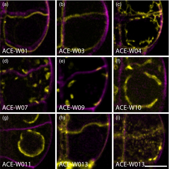
Arabidopsis cellular markers for embryogenesis driven by pWOX2 (ACE‐W) markers label cellular compartments in embryos.
Single optical sections of plasma membrane (a; ACE‐W01; AtPIP2A:GFP), inner membrane (b; ACE‐W03; BOR1:mCitrine), outer membrane (c; ACE‐W04; mCherry:NIP5;1), trans‐Golgi network and early endosomes (d; ACE‐W07; eYFP:VTI12), Golgi complex (e; ACE‐W09; eYFP:GOT1p), tonoplast and vacuole (f; ACE‐W10; eYFP:VAMP711), nuclear pore complex (g; ACE‐W11; AtNUP54:GFP) and plasmodesmata (h, i; ACE‐W13; mCherry:AtPDCB1) markers. Note that all markers are imaged in the center of one of the lower‐tier cells in an 8‐cell embryo, except panel (i), which is imaged at the upper cell surface. Scale bar for all panels = 5 μm. [Colour figure can be viewed at http://wileyonlinelibrary.com]
