Figure 7.
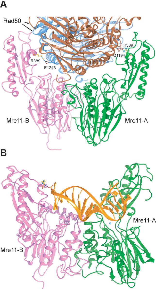
R389 at the Mre11–Rad50 and Mre11-DNA interfaces. (A) Residue R389 of Mre11 is localized at the interface between Mre11 and Rad50 in the ScMre11–Rad50 heterotetramer. The position of R389 (in yellow) is shown on each Mre11 monomer constituting a tetramer with Rad50 subunits (in light blue and brown). The interaction is not symmetric, involving different residues (E1243 or Q1194) on the different Rad50 subunits. (B) Interaction of R389 (in yellow) of dimeric ScMre11 (in pink and green) with dsDNA (in orange). Black line indicates the salt bridge between R389 and the phosphodiesterase bridge at the 3′ terminus of the dsDNA end. Mg2+ ions in the active site in one monomer are indicated in light blue.

