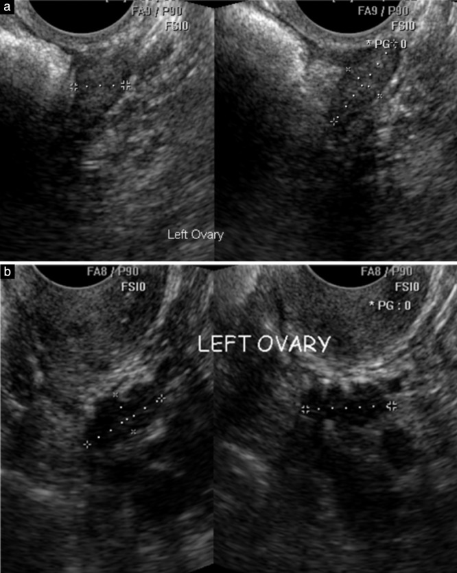
Figure 3.

Longitudinal and transverse transvaginal ultrasound images acquired by sonographers to measure left ovaries in postmenopausal women. (a) The expert judged the ovary as normal and correctly measured by the sonographer. (b) The expert considered the sonographer had mistakenly measured a section of bowel rather than the ovary as the haustrations of large bowel are clearly visible in the structure marked by the calipers.
