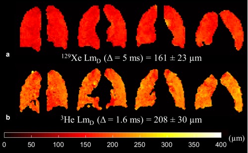
Figure 3.

Prospective CS results for a healthy volunteer (HV1) (SNR = 30). (a) Example 129Xe LmD maps derived from 3D multiple b‐value 129Xe DW‐MRI. (b) Example 3He LmD maps in comparative slices demonstrate the mismatch in LmD values between the two nuclei.
