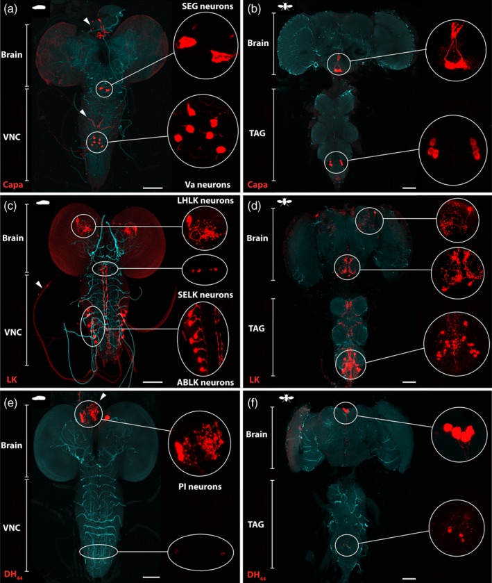
Figure 2.

Immunocytochemical localisation of Capa, LK and DH44 neuropeptides in the D. suzukii larval and adult central nervous system (CNS). (a, b) Localisation of Capa neurons using anti‐Capa precursor antibody in (a) the larval and (b) the adult CNS. Capa precursor immunoreactivity is localised to a single pair of very large neuroendocrine cells in the suboesophageal ganglion (SEG neurons) and three pairs of abdominal neuroendocrine cells (Va neurons). Arrowheads show neurohaemal organs, the retrocerebral complex for the two cell bodies in the suboesophageal neuromere, and the abdominal median nerves for the three pairs of ventral neuroendocrine cells in the abdominal neuromeres. (c, d) LK immunofluorescence using anti‐LK antibody in (c) the larval and (d) the adult CNS. LK‐expressing neurons are distributed as a pair of lateral horn LK (LHLK) neurons, two pairs of suboesophageal LK (SELK) neurons, and seven pairs in the abdominal ganglia of the larval CNS. In the ventral ganglion of the adult, around nine pairs of abdominal LK (ABLK) neurons have axons that leave the CNS by abdominal nerves and processes for the release of LK into the haemolymph. (e, f) Localisation of DH44 neurons using anti‐DH44 antibody in (e) the larval and (f) the adult CNS. DH44 peptides are produced in six neurons (three cells in each cerebral lobe) of the pars intercerebralis (PI), with axons running to the retrocerebral complex of the corpus cardiacum (arrow). Additional smaller set of neuroendocrine cells in the larval and adult ventral nerve cord are labelled with the DH44 antibody. All patterns of expression are representative of both males and females. Inserts are maximum projections of confocal z‐series showing magnifications of selected regions. VNC, ventral nerve cord; TAG, thoracic‐abdominal ganglion. Scale bars = 100 µm.
