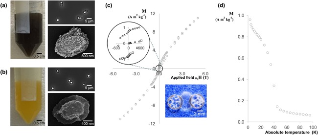
Figure 5.

Magnetic measurements and images depicting the color and appearance of B. megaterium spores that have undergone the adsorption procedure described in section 2.4.3. (a) manganese coated spores; (b) iron coated spores; (c) field dependency measurement for manganese coated spores at 5 K; the image shows a sample of spores aligned along the magnetic field lines between two circular neodymium magnets; (d) temperature dependency measurement for manganese coated spores with a magnetic field strength of 1,000 Oe. In (a) and (b) the images show: left, color of the spore suspension; top right, phase contrast image of the spores; bottom right, SEM image of the spore. Error bars indistinguishable when plotted
