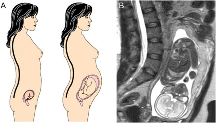
Figure 1.
(a) Schematic representation of increasing lumbar lordosis during pregnancy to compensate for fetal load and maintain a stable position of the center of mass (black and white circle with crosshairs). Adapted from (28). (b) MRI image of a 30-year-old woman at 25 weeks’ gestation showing marked lordosis. Figure 1a adapted with permission from Macmillan Publishers Ltd: Whitcome KA, Shapiro LJ, Lieberman DE. Fetal load and evolution of lumbar lordosis in bipedal hominins. Nature. 2007; 450:7172.

