Figure 5.
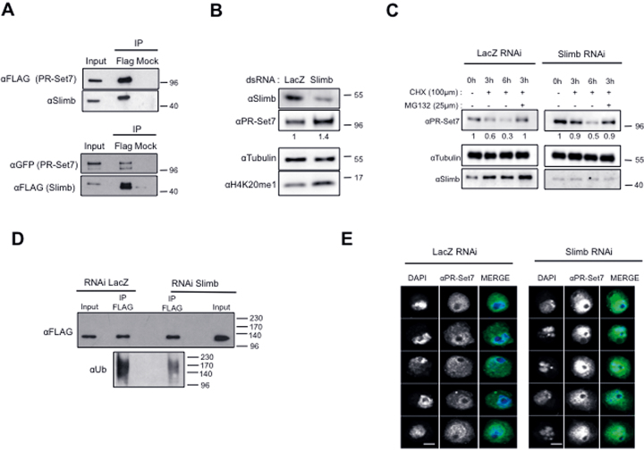
the SCFSlimb complex targets the nuclear pool of PR-Set7. (A) Immunoblot analysis for the indicated antibodies of immunoprecipitates (IP) done from lysates of S2 cells expressing FLAG-PR-Set7 (upper panel) or GFP-PR-Set7 and FLAG-Slimb (lower panel) using FLAG specific or irrelevant (Mock) antibodies. Input lane corresponds to 10% of cell lysates used per IP. (B) Immunoblot analysis for the indicated antibodies of whole cell extracts derived from S2 cells treated with LacZ as a negative control or Slimb specific dsRNA as indicated. Antibodies used are indicated on the left. (C) Immunoblot analysis of whole cell extracts derived from S2 cells incubated with LacZ (control, left) or Slimb dsRNA (right) during 5 days and treated with cycloheximide (CHX) alone or in combination with MG132 as indicated. Antibodies used are indicated in the left. Relative amounts of PR-Set7 normalized to tubulin in each RNAi condition are indicated. (D) Immunoblot analysis of immunoprecipitates (IP) done from lysates of S2 cells expressing ubiquitously (actin promoter) FLAG-PR-Set7 using αFLAG coupled or protein G coupled (Mock) dynabeads revealed with α-FLAG (top) and α-Ubiquitin (bottom) specific antibodies upon LacZ (control, left) or Slimb RNAi treatment during 5 days. Input lane corresponds to 10% of lysates used per IP. Note that 10% of the IP material was loaded on gel to detect FLAG protein. (E) Representative examples of S2 cells incubated with LacZ (left panels) or Slimb dsRNA during 4 days stained with PR-Set7 antibody (green) and DAPI (gray) and analyzed by immunofluorescence. Scale bars represent 3μm.

