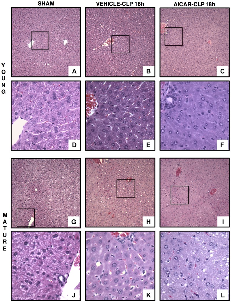
Figure 1.
Representative histology photomicrographs of liver sections. Normal liver architecture in control young [A, D (inset)] and mature [G, J (inset)] mice. Liver damage in vehicle-treated young (B) and mature (H) mice at 18 h after CLP with edema, inflammatory cell infiltration, and lipid vacuoles [E, K (insets)]. Amelioration of liver architecture in AICAR-treated young mice [(C, F (inset)]. Amelioration of liver architecture in AICAR-treated mature mice (I) with presence of small lipid vacuoles (L, inset) (n = 4–6 different tissue sections in each experimental group showed similar patterns). Original magnification: ×100 (A–C, G–I); ×400 (D–F, J–L).

