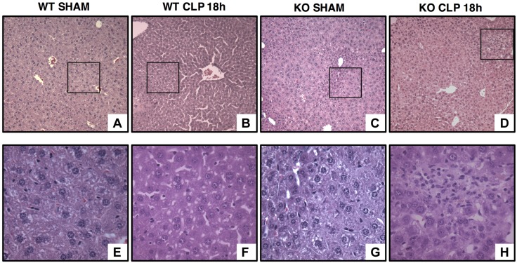
Figure 3.
Representative histology photomicrographs of liver sections of young AMPKα1 WT and KO mice. Normal liver architecture in young WT (A, inset shown in E) and KO mice (C, inset shown in G). Liver damage in young WT (B) and KO (D) mice after sepsis with edema, necrosis, and inflammatory cell infiltration [F, H (insets)] (n = 4 different tissue sections in each experimental group showing a similar pattern). Original magnification: ×100 (A–D); ×400 (E–H).

