Figure 3.
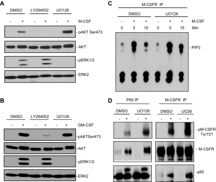
Blocking ERK activation increases M-CSF–evoked M-CSFR–Tyr721 phosphorylation and PI3K activation. A, B) WT BMMs were starved; pretreated with the MEK inhibitor UO126 (10 µM), the PI3K/mTOR inhibitor LY294002 (10 µM), or vehicle (DMSO) for 1 h; and then were either left unstimulated (−) or stimulated (+) for 10 min with 30 ng/ml M-CSF (A) or 50 ng/ml GM-CSF (B). AKT and ERK activation were assessed in TCLs (30 µg) by immunoblotting with the indicated p-specific antibodies. Blocking ERK activation by MEK inhibitor treatment increases M-CSF-, but not GM-CSF–evoked AKT activation, whereas inhibiting M-CSF or GM-CSF–evoked PI3K activation did not affect ERK activation. C, D) MEK inhibitor treatment increases M-CSFR–associated PI3K activity and M-CSFR Tyr721 phosphorylation and association with the p85 subunit of PI3K. WT BMMs were starved, pretreated for 1 h with vehicle (DMSO), UO126 (10 µM), or LY294002 (10 µM); and then stimulated with M-CSF (30 ng/ml) for the indicated times. M-CSFR immunoprecipitates (from 1 mg TCL) were subjected to PI3K assays (C). M-CSFR and p85 immunoprecipitates were also immunoblotted with the indicated antibodies (D). Data shown are representative of the 3 independent experiments.

