Figure 6.
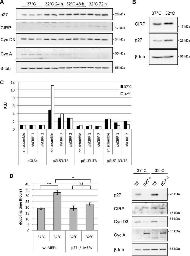
CIRP induces p27 translation in mild hypothermia. (A) MEFs were grown at 37°C (control, normothermia) or incubated at 32°C (mild hypothermia) for the times indicated. The expression of p27, CIRP, cyclin D3, cyclin A and β-tubulin was determined by western blot. (B) HEK293 cells were moved to 32°C and incubated for 40 h. Protein extracts from these cells or cells grown at 37°C were subjected to immunoblot analysis for CIRP, p27 and β-tubulin. (C) HEK293 cells were transfected with CIRP shRNA or scramble shRNA expression plasmids, luciferase reporter plasmids (depicted in Figure 2A) and Renilla luciferase for normalization control. 25 h after transfection, one aliquot of cells was incubated at 32°C for additional 25 h. Relative luciferase activity (RLU) was determined as ratio of firefly and Renilla luciferase activities and normalized for the activity of pGL3-control. One representative experiment out of four is shown. (D) Average doubling time of wt MEFs and p27−/− MEFs grown at 37 or 32°C. Wt MEFs and p27−/− MEFs were seeded and their proliferation was followed by counting the cells at timepoints starting 5–10 h after shifting to the lower temperature incubator and for 72 h. Cell numbers obtained during the exponential growth phase were used to calculate the doubling time. Data are shown as mean ± SEM, n = 5 independent experiments. Unpaired two-tailed t-test was used to compare the average doubling time of MEFs at 37°C and at 32°C and of p27−/− MEFs at 37°C and at 32°C; *P < 0.05, **P < 0.01, ***P < 0.001. The difference between the average doubling time of MEFs at 32°C and p27−/− MEFs at 32°C is also statistically significant, based on the same test. A representative immunoblot analysis of these cells is shown (right panels). Cells were collected after 8 h at 32°C and the expression of p27, CIRP, cyclin D3, cyclin A and β-tubulin was determined.

