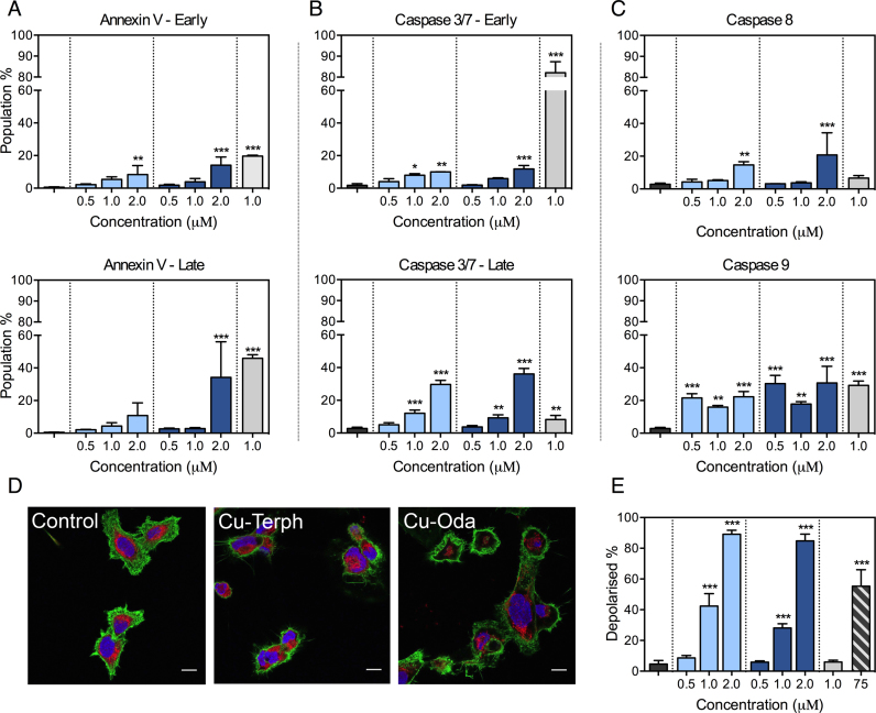
Figure 2.
SKOV3 cells are treated over 24 h with varying concentration of Cu-Oda and Cu-Terph (0.5–2.0 μM) and control agents Dox (1.0 μM) and CCCP (75 μM). (A) Populations of early apoptotic cells exhibiting positive staining for Annexin V only with late apoptotic population detected through positive staining for both Annexin V and 7-AAD. (B) Detection of caspase 3/7 in the absences or presence of 7-AAD positive staining indicative of mid- and late-apoptotic activation. (C) Activation of caspase 8 (extrinsic pathway) and caspase 9 (intrinsic pathway). (D) 100 × magnification of SKOV3 cells treated with di-nuclear copper complexes, Cu-Oda and Cu-Terph (1.0 μM). Nuclei are stained with DAPI, F-actin with Alexa Fluor 488-Phalloidin and mitochondria with MitoTracker Deep Red. Scale bar indicates 10 μm. Individual channels are shown in Supplementary Figure S4. (E) Population of depolarized mitochondria through the detection of potential-sensitive shift in JC-1 emission. Hypsochromic shifts, evident in scatter plot, demonstrates drug-induced depolarization (Supplementary Figure S5). Non-significant (ns) P > 0.05; *P ≤ 0.05; **P ≤ 0.01; ***P ≤ 0.001.

