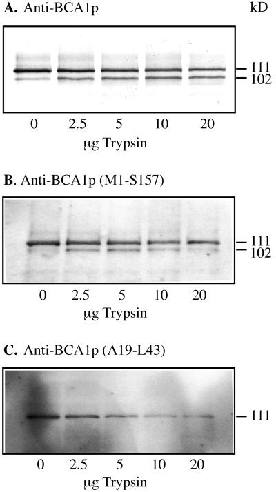
Figure 3.
Western analysis of BCA1p in low-density membranes after trypsin treatment, carried out as described in Askerlund (1996). Samples were collected from the complete Ca2+-pumping assay exactly 2 min after the addition of trypsin. The assay mix was trichloroacetic acid-precipitated, and analyzed by western blotting using anti-BCA1p (A), anti-BCA1p (Met-1 to Ser-157) (B), and anti-BCA1p (Ala-19 to Leu-43) (C). The lanes received 4 μg of protein.

