Figure 5.
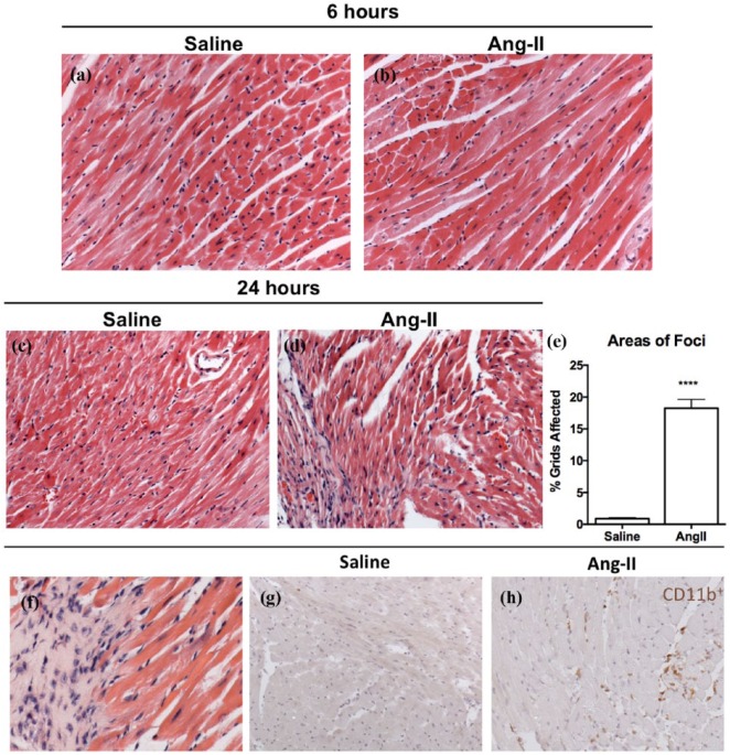
Mononuclear cellular infiltration in the myocardium was present after 24 hours of Ang-II infusion. Representative images of myocardial cross-sections stained with H&E from mice that received ((a) and (c)) saline or ((b), (d) and (f)) Ang-II. (e) H&E sections were semi-quantified for cellular infiltration in Ang-II animals relative to saline controls. Immunohistochemistry was used to characterize mononuclear cell infiltrates as CD11b+. Representative myocardial cross-sections from animals treated with (g) saline or (h) Ang-II.
Data are expressed as means ± SEM.
n = 5.
****p < 0.0001, compared with saline control. Original magnification: ×20 (a) to (d); ×40 (f).
Ang-II: Angiotensin II; H&E: hematoxylin and eosin.

