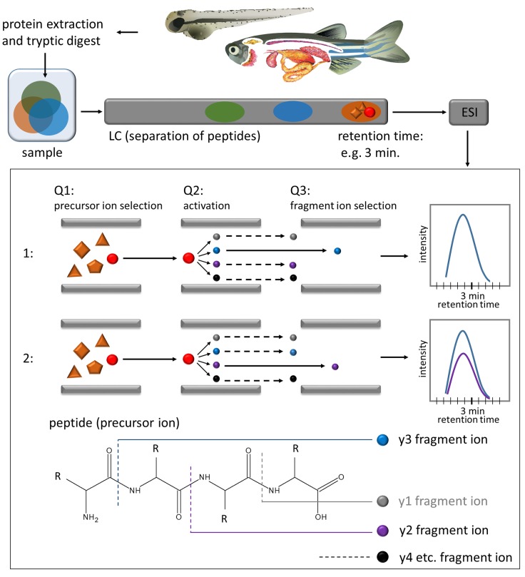
Figure 1.
Overview of experimental workflow and schematic representation of the MRM technique. The proteins are extracted from tissue, tryptically digested into peptides, separated with liquid chromatography (LC), ionized via electrospray ionization (ESI) and transferred into the triple-quadrupole mass spectrometer (Q1–Q3). In Q1—the precursor ion (peptide) is selected, in Q2 the peptide is fragmented into fragment ions (y1, y2. y3, etc.), in Q3 the selection of the fragment ion takes place. Subsequently, the intensity of the fragment ion is measured over time. Within 1 measurement, several transitions (peptide and fragment ion) can be monitored.

