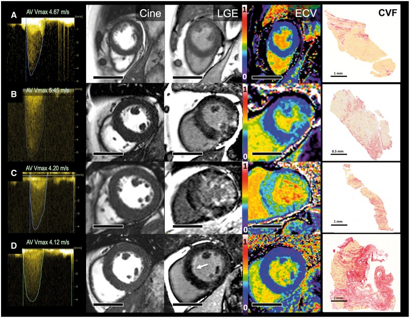
Figure 1.
Aortic stenosis, myocardial hypertrophy and fibrosis by imaging and biopsy. Four exemplar patients showing continuous-wave Doppler (maximum velocities >4m/s; Column 1), short axis cine stills demonstrating degrees of left ventricular hypertrophy (Cine; Column 2), matching late gadolinium enhancement images (LGE, Column 3), matching extracellular volume fraction (ECV, Column 4), myocardial biopsy stained with picrosirus red [collagen volume fraction (CVF), Column 5]. Patient A has minimal LVH, no LGE, an ECV of 28.4% and minimal biopsy subendocardial fibrosis (CVF 4.6%). Patient B has concentric LVH, patchy non-infarct LGE, an ECV of 29.9% and moderate biopsy fibrosis (CVF 19.3%). Patient C has concentric LVH, widespread non-infarct LGE, an ECV of 36.5%, and severe biopsy fibrosis (CVF 24.5%). Patient D has mild concentric LVH, subtle subendocardial LGE (white arrow), an ECV of 24.5%, thickened endocardium, and subendocardial scarring. Scale bars (Columns 2–4) equal 5 cm.

