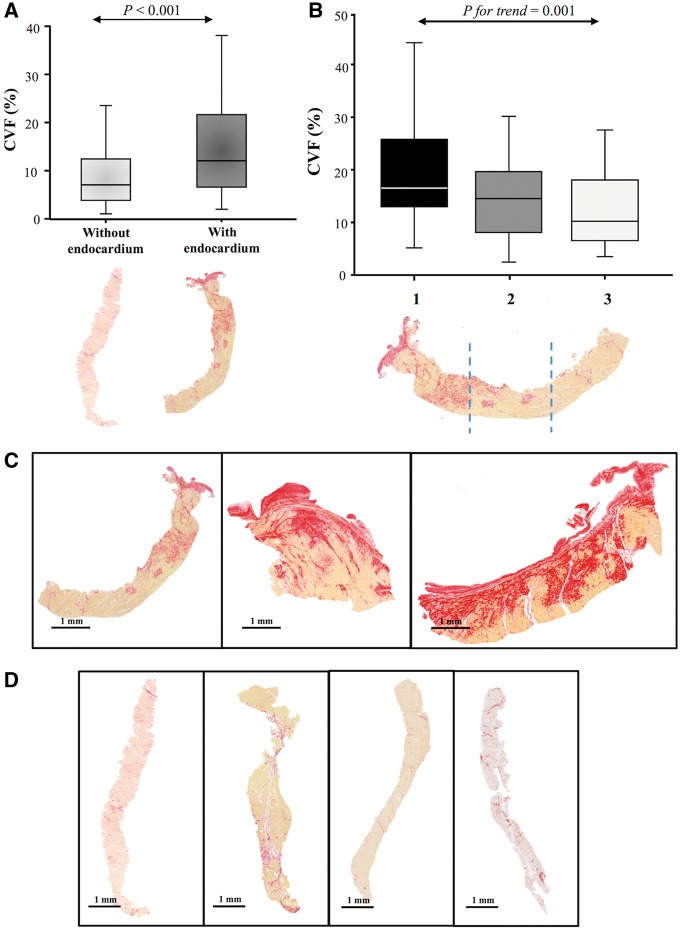
Figure 2.
Biopsies with and without endocardium—presence of a gradient of fibrosis. (A) Collagen volume fraction in biopsies with and without endocardium. (B) Collagen volume fraction in samples with endocardium divided in tertiles. Box plots show the 5th and 95th (vertical lines), 25th and 75th (boxes), and 50th (horizontal line) percentile values for collagen volume fraction. (C) Representative images of three biopsies with endocardium (left panel needle and middle and right panel scalpel). (D) Representative images of four biopsies without endocardium (needle).

