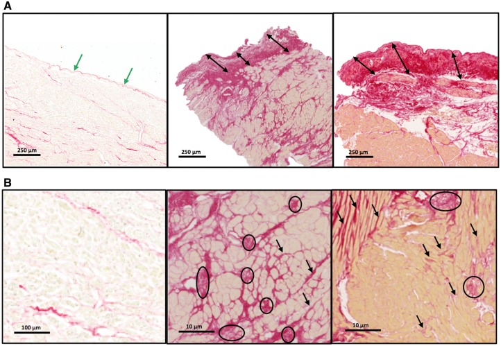
Figure 3.
Patterns of fibrosis—endocardial thickening and myocardial microscars and interstitial fibrosis. (A) The endocardium in a control subject (left panel) and in two aortic stenosis patients at the same magnification. The arrows show the endocardial thickness (green in normal, black in aortic stenosis). (B) Higher magnification showing minimal subendocardial interstitial fibrosis in a control subject (left panel) and extensive microscars and interstitial fibrosis in two aortic stenosis patients. Circles identify microscars and arrows diffuse fibrosis.

