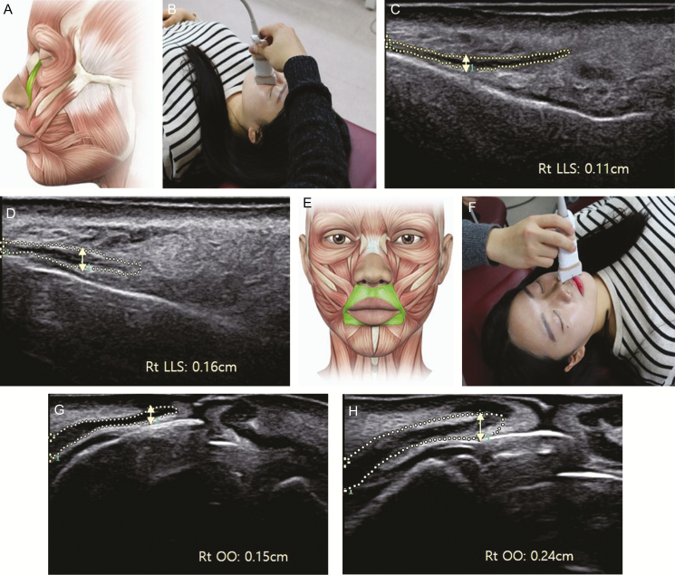
Figure 2.
Measurement of the thickness of the levator labii superioris and orbicularis oris muscles using ultrasound images of a 30-year-old woman. (A) Anatomical position of the levator labii superioris muscle, (B) probe position for the levator labii superioris and muscle thickness of the levator labii superioris (C) initially and (D) after the sessions; (E) anatomical position of the orbicularis oris muscle, (F) probe position for the orbicularis oris and muscle thickness of orbicularis oris (G) initially, and (H) after the sessions.

