Figure 6.
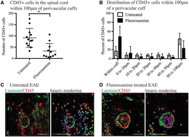
Targeting CSPGs with fluorosamine reduces the infiltration of CD45+ cells into the spinal cord parenchyma. (A) CD45+ cells were stained by immunohistochemistry and rendered as ‘spots’ in Imaris. Imaris quantified the number of CD45+ cells in the spinal cord 100 µm from perivascular cuffs (identified with pan-laminin staining). CD45+ cells within the perivascular cuff were not included in this analysis. (B) Distribution of CD45+ cells within 100 µm of perivascular cuffs in fluorosamine-treated and untreated EAE spinal cords. (C) Left, immunohistochemistry of a perivascular cuff in an untreated mouse and right, Imaris rendering of laminin staining as a surface and CD45+ as spots overlain on true CD45+ (red) staining. Distance of CD45+ cells from the perivascular cuff is represented in their colour gradient (shown in the bottom left), with purple representing nearby and red far. (D) Left, immunohistochemistry of a perivascular cuff of fluorosamine-treated EAE mouse. Right, Imaris rendering of laminin as a surface and CD45+ as coloured spots, overlain on true CD45+ (red) staining. Distance of CD45+ cells from the perivascular cuff is represented in their colour gradient (shown in the bottom left), with purple representing nearby and red far. Spinal cords from four mice in each group were quantified. Scale bars = 50 μm.

