Figure 1.
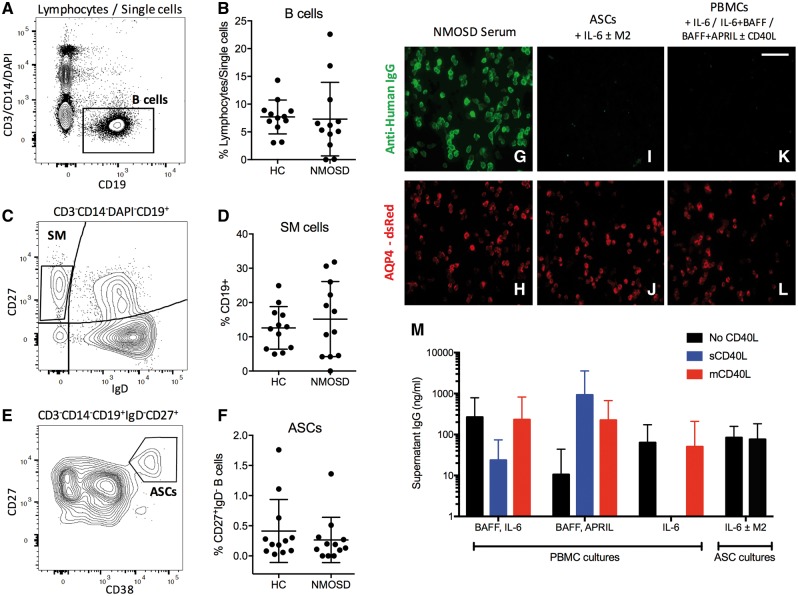
B cell flow cytometry from patients with AQP4-IgG positive NMOSD and healthy controls. Ex vivo PBMCs from patients and healthy control subjects (HCs) gated as single CD3−CD14−DAPI−CD19+ B lymphocytes (A and B), CD27+IgD− switched memory cells from the B cell gate (SM; C and D) and CD27++CD38++ (ASCs; E and F) from the CD27+IgD− gate. As expected, NMOSD patient serum IgG bound the surface of AQP4 and dsRed co-expressing cells (G and H, magnification ×400). After 13 days in culture, AQP4-IgG was not detected from sorted patient ASCs [cultured with IL-6 and/or the bone marrow stromal cell line, M2-10B4 (M2); I and J] or from PBMCs cultured under ASC-maintenance conditions (IL-6; BAFF with IL-6; BAFF with APRIL, all ± CD40L; K and L). Total IgG determination from PBMC and ASC cultures from 12 patients was comparable to literature values (M). Scale bar = 100 µm.

