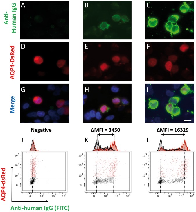
Figure 3.
Determination of AQP4-IgG from serum and culture supernatants. Binding of patient IgG from culture supernatants (A–C; green, magnification ×1000; Scale bar = 10 µm) to live HEK-293T cells co-expressing AQP4 and dsRed from a single plasmid (D–F). Merge shown with DAPI (G–I). Sera or supernatants found to be positive by this cell-based assay, underwent flow cytometry to generate quantitative titres, defined as the difference in MFI between the dsRed-expressing (red) and dsRed-negative (black) cell populations (expressed as ΔMFI, J–L). Examples of negative (A, D, G and J), moderately (B, E, H and K) and strongly (C, F, I and L) positive samples shown.

