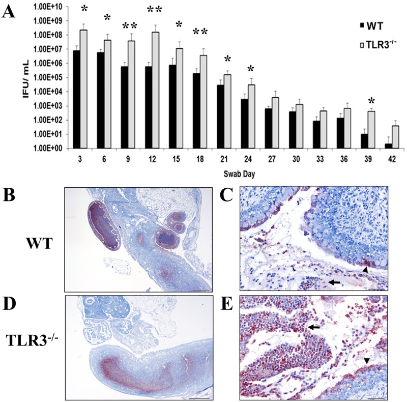
Fig 1. The levels of Chlamydia muridarum shedding from genital tracts of TLR3-/- mice are significantly higher during the first 24 days of infection.
A) Genital tract infections were performed, swabs were collected every third day for 42 days, and C. muridarum titers were determined by infecting fresh McCoy cell monolayers (see Materials and methods). Representative data from an average of 5 mice are shown. IFU = inclusion forming units. * = p <0.05; ** = p <0.005. B-E) Immunostaining of Chlamydia muridarum in genital tracts to wild-type (WT) and TLR3-/- mice at day 7 post-challenge. Detection of Chlamydia antigen (red staining) within neutrophils (arrow) and epithelial cells (arrowhead) in oviducts (B), endometrium (C), and cervix from a representative WT mouse. Detection of Chlamydia antigen (red staining) associated with neutrophils (arrows) and epithelial cells (arrowhead) in endometrium and cervix (D and E), from a representative TLR3-/- mouse.

