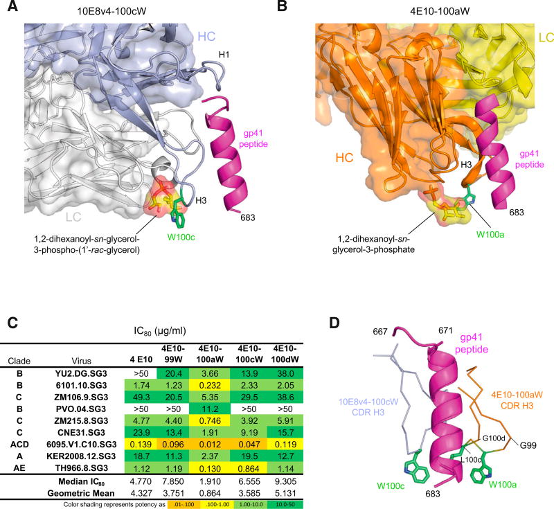
Figure 4. MPER-Directed Antibody 4E10 Shares the Membrane-Binding Functional Hotspot of 10E8.
(A) Structural model of antibody 10E8v4 recognizing lipid headgroups, as defined by Irimia et al. (2017), with hotspot position 100c shown in green.
(B) Structural model of antibody 4E10 recognizing lipid headgroups, as defined by Irimia et al. (2016).
(C) Virus neutralization by 4E10 and variants showing 100aW with improved activity.
(D) Superposition gp41 peptide as recognized by 4E10 and 10E8 identifies 4E10 CDR H3 as a hot-spot of functional enhancement.

