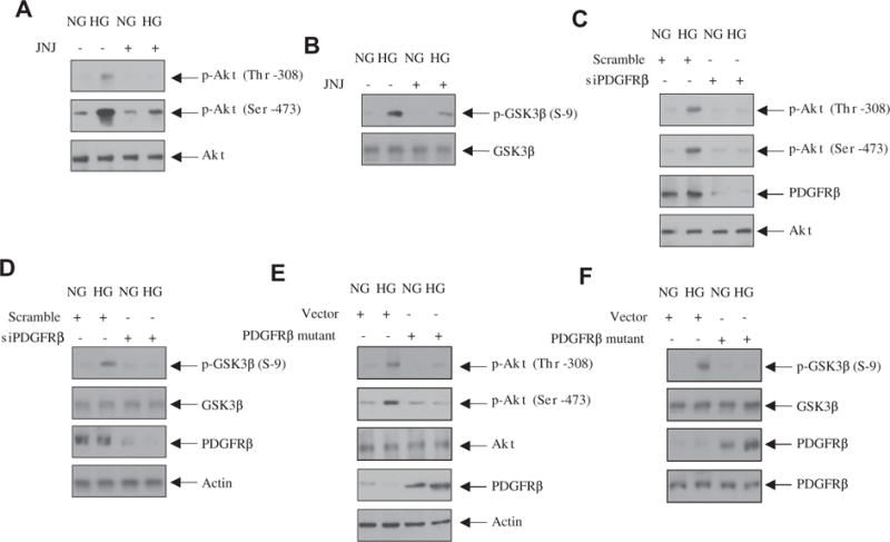
Fig. 2.

PDGFRβ regulates high glucose-stimulated Akt kinase activity. (A and B) Mesangial cells were treated with 0.1 μM JNJ for 1 h prior to incubation with high glucose (HG) or normal glucose (NG) for 24 h. Equal amounts of cell lysates were immunoblotted with the indicated antibodies. (C–F) Mesangial cells were transfected with siPDGFRβ (panels C and D) or with PDGFRβ Y740/751F (E and F) mutant. The cells were then incubated with high glucose (HG) or normal glucose (NG) for 24 h. The cell lysates were immunoblotted with indicated antibodies.
