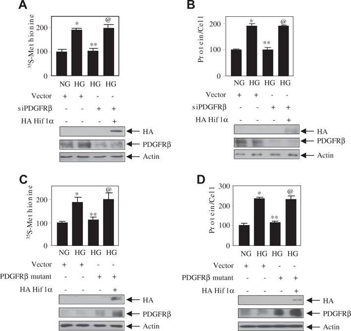
Fig. 6.

Hif1α downstream of PDGFRβ controls high glucose-induced mesangial cell hypertrophy. Mesangial cells were transfected with siPDGFRβ or PDGFRβ mutant (Y740/751F) along with HA Hif1α. The transfected cells were incubated with normal glucose or high glucose. The protein synthesis and hypertrophy were determined as described in the Materials and methods section [22,25]. The bottom panels show the expression of HA Hif1α, PDGFRβ and actin. Mean ± SE of 3–6 measurement is shown. *p < 0. 01, 0.05 or 0.001 vs NG; **p < 0. 01, 0.05 or 0.001 vs HG; @p < 0.05, 0.01 or 0.001.
