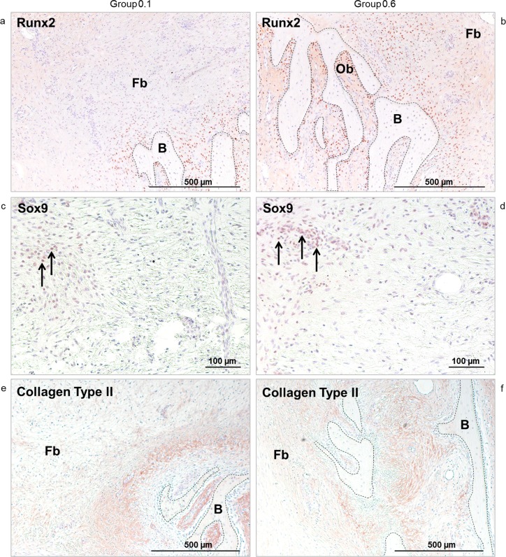
Fig 6.
a-f. Immunohistochemistry. The intensive red to brown coloring indicates osteoblasts (Ob) and fibroblast-like cells staining positive for Runx2 (Fig 6A and 6B). Osteo-chondroprogenitor cells in the zone adjacent to the titanium plate surface are positive for Sox9 (arrows) (Fig 6C and 6d). Collagen Type II positive areas are visible as red to brown coloring in the collagenous matrix surrounding newly formed bone (B) but not in the fibrous tissue (Fb) distant to it (Fig 6E and 6F). Pictures on the left represent Group 0.1, pictures on the right represent Group 0.6. 50-fold (a-b, e-f) and 100-fold (c-d) magnification.

