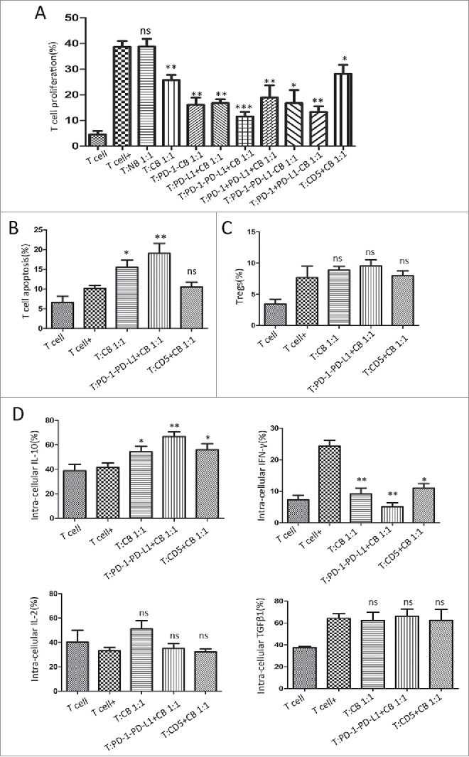
Figure 5.

PD-1−PD-L1+CD19+ B cells display immuno-regulatory properties on activated T cells. PD-1−PD-L1+CD19+ B cells were isolated from the splenic cells of tumor-bearing mice. T cells were cultured for 2 days with or without B cells for the subsequent analyses. (A) Regulation of CD3+ T-cell proliferation by different B cell subsets is shown in relative to T cells cultured in the absence of B cells from 8 independent experiments. (B) The apoptosis of CD3+ T cell was detected by FC at 48 h following incubation with splenic CD19+ cells, PD-1−PD-L1+CD19+cells or CD5+B cells from tumor-bearing mice. (C) The percentage of Tregs (CD4+CD25+CD125low) with or without B cell incubation was evaluated by FC analysis. (D) Cytokine concentrations (IL-10, IFN-γ, TGFβ1, and IL-2) were determined by FC to analyze T-cell intra-cellular production. Data were collected from 5 independent experiments. Data represent the mean ± SEM of 5 independent experiments. * = P < 0.05; ** = P < 0.01; *** = P < 0.001, ns = not significant as determined with t test or One-way ANOVA. BrdU, bromodeoxyuridine; MDSCs, myeloid-derived suppressor cells; Tregs, regulatory T cells; FC, flow cytometry; TNF, tumor necrosis factor; TGF, transforming growth factor; IL, interleukin; IFN, interferon.
