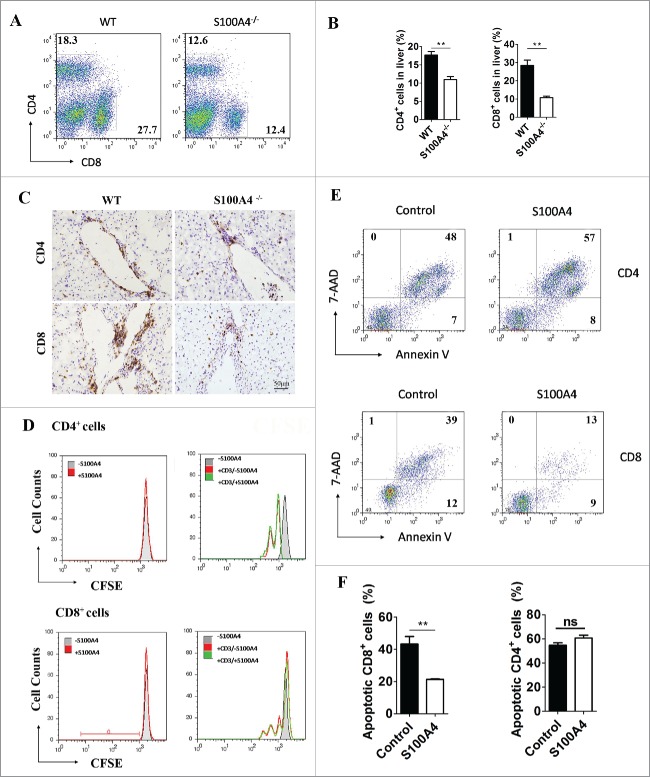
Figure 5.
S100A4 potentiates CD8+ T cell survival. (A) Representative FACS data and (B) quantification of CD4+ T cells and CD8+ T cells in the liver of WT or S100A4−/− mice (treated with 2A weekly for 4 weeks) were detected by FACS, **p < 0.01. (C) IHC staining for CD4+ and CD8+ in the liver tissue of WT and S100A4−/− mice. Scale bar, 50 μm. (D) CFSE-labeled CD4+ T and CD8+ T cells from the spleen were left unstimulated (left) or stimulated with anti-CD3 antibody (right) with or without soluble S100A4. T cell proliferation was analyzed by CFSE dilution. (E) and (F) CD4+ T and CD8+ T cells from spleen cells were stained with 7-AAD and Annexin V, and cells apoptosis was detected by FACS, **p < 0.01.

