
Figure 4.
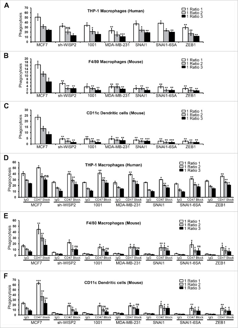
EMT upregulated CD47 inhibits the phagocytosis of EMT-activated cells and increased phagocytosis of human breast cancer cells after CD47 blockade.(A) EMT-activated cells and epithelial MCF7 cells were stained with CFSE, co-cultured with Human THP-1 derived macrophages for 2 h (stained with CellTrace™ Far Red-APC) at different target to effector (cancer: macrophage) ratios, and analyzed by flow cytometry. Phagocytosis is shown as the percentage of CFSE+APC+ phagocytosed cancer cells. The experiment was repeated 6 times. (B and C) EMT-activated cells and epithelial MCF7 cells were stained with CFSE, co-cultured with mouse macrophages or dendritic cells for 2 h at different target to effector (cancer: macrophage or dendritic cells) ratios, stained with F4/80-APC or CD11c-APC antibody and analyzed by flow cytometry. Phagocytosis is shown as the percentage of CFSE+APC+ phagocytosed cancer cells. The experiment was repeated 3 times. (D-F) Phagocytosis assay was performed with EMT-activated cells and epithelial MCF7 cells as described in 4A in the presence of CD47 blocking antibody (B6H12; e Bioscience) or matching IgG control by using Human THP-1 derived macrophages (D), mouse macrophages (E) or mouse dendritic cells (F). The experiment was repeated 3 times.