Figure 3.

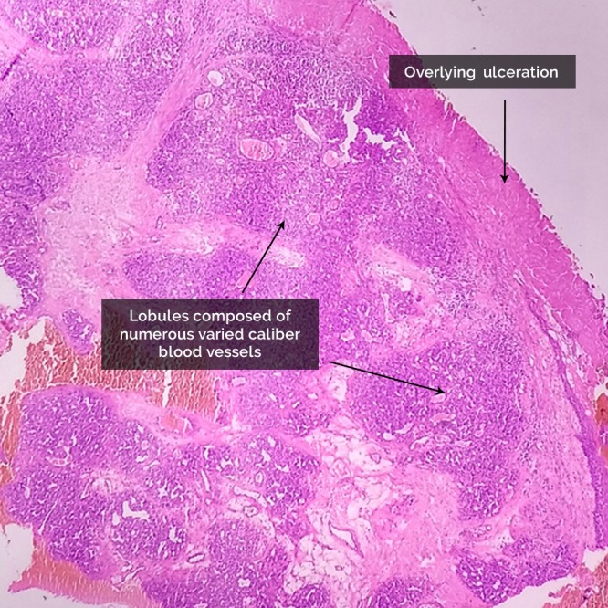
Histopathological examination showing lobules composed of numerous varied caliber blood vessels with an overlying ulceration. (Hematoxylin and eosin stain, ×10).

Histopathological examination showing lobules composed of numerous varied caliber blood vessels with an overlying ulceration. (Hematoxylin and eosin stain, ×10).