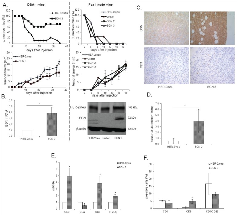
Figure 4.

Reduced tumor growth of BGN transfectants in vivo in immune competent mice. A. Frequency and diameter of tumors 1 × 10⁶ BGNlow/neg and BGNhigh HER-2/neu+ cells/mouse were subcutaneously injected as described in Material and Methods. Tumor growth was monitored overtime regarding frequency and diameter of the tumors. B. mRNA and/or protein expression of HER-2/neu and BGN in BGNlow/neg vs. BGNhigh HER-2/neu+ tumors. The mRNA and/or protein expression of HER-2/neu and BGN was determined in theBGNlow/neg and BGNhigh tumors as described in Materials and Methods using qPCRand Western blot analysis. C. Immunohistochemical staining of BGN and CD3 in BGNlow/neg and BGNhigh HER-2/neu+ cells. IHC of tumors was performed as described in Materials and Methods using anti- BGN, anti-HER-2/neu and anti-CD3 specific mAbs, respectively. D. Analysis of the number of CD3+ immune cells. The number of CD3+ immune cells was determined upon staining with an anti-CD3 mAb at 400 x magnification (HPF) considering preferentially areas with higher intra-tumoral immune cell density. E. Determination of the mRNA expression of immune markers and MHC class I expression in BGNlow/neg and BGNhigh HER-2/neu+ cells. MRNA expression of the different immune cell markers CD3, CD4, CD8, IL-2, FoxP3 and the MHC class I HC was determined by qPCR as described in Materials and Methods. The expression levels of these markers were compared in BGNlow/neg vs. BGNhigh HER-2/neu+ tumor lesions. F. Immune marker expression in peripheral blood of tumor bearing mice. The expression of levels of percentages of CD4, CD8 and CD4/CD25 were determined after injection of BGNlow vs. BGNhigh HER-2/neu+ tumor cells in the peripheral blood of mice using flow cytometry as described in Materials and Methods.
