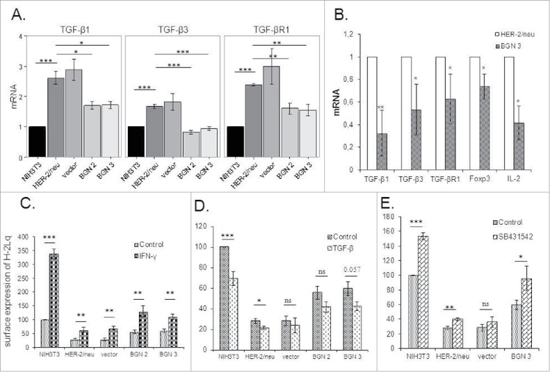
Figure 5.

Changes of the TGF-β pathway and MHC class I surface antigens by treatment with TGF-β and IFN-γ. A. mRNA expression of TGF-β isoforms 1 (TGF-β1 and 3) and the TGF-βR1 in BGNlow/neg and BGNhigh HER-2/neu+ cells. The expression of components of the TGF-β pathway was analysed by qPCR as determined in Materials and Methods. The results represent the mean of three independent experiments and expressed relative to NIH3T3 cells (set 1). B. Analysis of TGF-β and Treg in BGNlow vs. BGNhigh HER-2/neu+ cells. The expression levels of TGF-β isoforms (TGF-β1 and 3) and receptor (TGF-βR1), were analysed in BGNhigh HER-2/neu+ tumor lesions in vivo using qPCR. C. Enhanced MHC class I surface expression upon IFN-γ treatment. Untreated and 20ng/ml IFN-γ-treated cells were subjected to flow cytometry as described in Materials and Methods. MFI was determined and expressed relative to NIH3T3 cells (100%). D. Effects of TGF-β on MHC class I surface expression. Untreated and 40ng/ml TGF-β-treated cells were subjected to flow cytometry as described in Materials and Methods. E. Influence of the TGF-β inhibitor on MHC class I surface expression. NIH3T3, BGNlow/neg and BGNhigh HER-2/neu+ cells were either left untreated or treated with 20 ng/ml TGF-β inhibitor (SB431542), before MHC class I surface expression was determined by flow cytometry. MFI of MHC class I surface expression of untreated and SB431542-treated BGNlow/neg and BGNhigh HER-2/neu+ cells was correlated to MFI of untreated NIH3T3 cells, which was set 100%. The experiments were at least performed three times and results represent the mean of these experiments.
