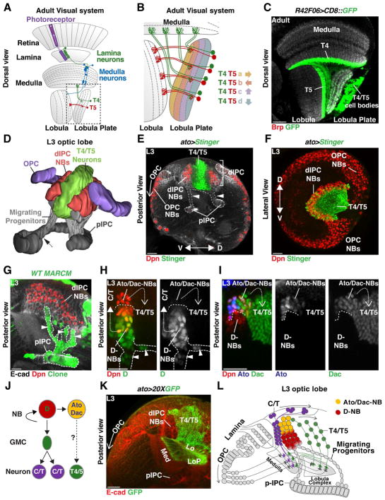
Figure 1. Development of the dIPC.
(A) Schematic of an adult optic lobe highlighting the ON (T4) and OFF (T5) pathways of the motion detection circuit. Visual input propagates retinotopically via T4 and T5 neurons to the four layers of the Lobula Plate. Dashed line shows detail in panel (B).
(B) Four subtypes of T4 and of T5 neurons, each directionally tuned to one of the four cardinal directions, converge onto layers a and b of the horizontal system, or layers c and d of the vertical system in the Lobula Plate.
(C) Dorsal cross-section of an adult brain in which R42F06-Gal4 (T4/T5-Gal4) drives membrane-GFP (green) expression in T4 and T5 neurons. T4 dendrites project to the Medulla and T5 to the Lobula (neuropiles labeled in grey by anti-Brp). (A, B, C) anterior is to the left.
(D) Posterior view of a 3-D model of the developing larval Outer (purple) and Inner (grey) Proliferation Centers (OPC and IPC). Dashed arrow marks the boundary between the proximal-IPC (pIPC) and the surface IPC. Progenitors delaminate from the pIPC (grey) to become neuroblasts (NBs) in the distal-IPC (dIPC) (red). T4 and T5 neurons (green) are generated by dIPC NBs and form inside the dIPC crescent. See also Movie 1.
(E, F) Red-Stinger driven by Ato-Gal4 labels T4/T5 neurons.
(E) Posterior cross- section of a developing larval optic lobe. The pIPC-neuroepithelium is localized between the OPC and the central brain. Cells delaminating from the pIPC (arrowheads) migrate to form the dIPC-NBs (Dpn, red) that will generate T4 and T5 neurons (green). In the OPC, a proneural wave converts the OPC-neuroepithelium into Medulla NBs (Dpn, red) from proximal to distal (open arrow).
(F) dIPC-NBs form a U-shaped cluster (Dpn, red) along the dorsal-ventral axis that surrounds T4 and T5 neurons (green). Anterior is to the left.
(G) pIPC-MARCM clone (green). Migrating cells (arrowheads) delaminate from specific neuroepithelial locations and form dIPC NBs (Dpn, red) that spread along the dorso-ventral axis (see (F) and Movie 1).
(H,I,J) dIPC-NBs transit trough two temporal stages: Migrating cells (arrowheads) and newly produced NBs express Dichaete (D) [green in (H)] that produce distal C/T neurons (closed dashed arrow); Older NBs express Ato [blue in (I)] and Dac (green) and produce Dac+ T4/T5 neurons (open dashed arrow).
(J) Schematic summarizing neurogenesis of the dIPC.
(K) T4/T5 neurons project to their target Medulla, Lobula or Lobula Plate (Ecad, red) neuropiles during development.
(L) Schematic summarizing the development and neurogenesis of the dIPC. In this and subsequent figures scale bar is 20 μm.

