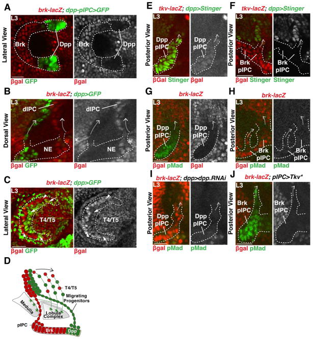
Figure 2. IPC-derived migrating progenitors populate the dIPC.
(A) Lateral view of the pIPC-neuroepithelial Brk domain marked by brk-lacZ (red) flanked by Dpp domains marked by dpp-pIPC>GFP (green). Anterior is to the left.
(B) Chains of migrating progenitors (arrows) emerging from the Dpp (green) or Brk domains (red).
(C) Lateral view of the dIPC. Anterior is to the left. Dpp-derived NBs (arrows, green) and Brk-derived NBs (arrowheads, red) spread equally within the dIPC. T4/T5 neurons are located in the inside of the crescent.
(D) Schematic summarizing the development of the dIPC.
(E,F) The Dpp receptor Tkv is expressed in both the Dpp (E) and Brk domains (F).
(G,H) Dpp signalling was active (pMad) in the Dpp (G), but not in the Brk domain (H).
(I) Expression of dpp-RNAi in the Dpp domains lead to the expansion of brk-lacZ expression in the Dpp domain and loss of pMad.
(J) Expression of an activated form of Tkv (Tkv*) downregulated brk-lacZ and leads to ectopic pMad in the Brk domain.

