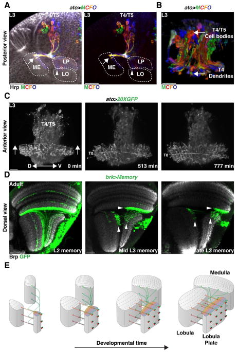
Figure 7. Neurogenesis mode and neuronal birth location establish retinotopy.
(A,B) In a developing L3 larval brain, T4/T5 neurons projected to the neuropiles along the dorso-ventral axis according to their birth location.
(A) Posterior view of MCFO labeled T4/T5 neurons. Ventral is to the left.
(B) Anterior 3-D view of MCFO labeled T4/T5 neurons. Proximal is to the bottom. Neurons projected in the neuropiles along the dorsal-ventral axis according to their birth position. Ventrally born T4/T5 neurons projected ventrally in the neuropiles [arrow in (A)]. See also Movie 5.
(C) Still pictures of a time-lapse movie highlighting T4 projections in the Medulla. Anterior view with proximal to the bottom. Neurons innervating columns along the same dorso-ventral plane projected at the same time in the neuropiles. Dashed line follows the position of neurons that were targeting the medulla at T0 (see Movie 6). Arrows indicate the development along the anterior-posterior axis of the Medulla neuropile.
(D) Activation of a memory cassette in the Brk-pIPC domain at late L2, mid-L3 and Late L3 and visualized in the Adult with T4/T5-Gal4 labeled neurons of the horizontal system along the antero-posterior axis (Arrowheads) depending on their time of birth (see also Figure S7).
(E) 3-D Schematic illustrating the establishment of the retinotopy of T4 and T5 neurons.

