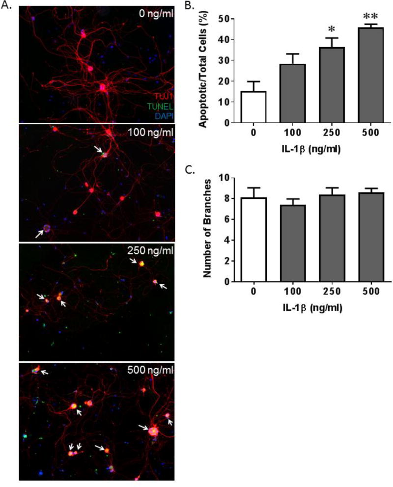
Figure 5.
IL-1β induces apoptosis and an increase in neurite length in pelvic neurons in vitro. A. Representative images of pelvic neurons stained for TUJ1 (red; neuron specific class III β-tubulin), TUNEL (green, apoptotic marker) and DAPI (blue; nuclear stain). Apoptotic neurons are indicated by the white arrows. B. The number of apoptotic pelvic neurons, normalized to total neurons, per area of interest in cultures treated with increasing concentrations of IL-1β. C. The number of branches of the neurons visualized for B. Data in all graphs are presented as the mean ± SEM. n=4 for all groups. * indicates p<0.05 and ** indicates p<0.01 versus the 0 ng/ml IL-1β sample.

