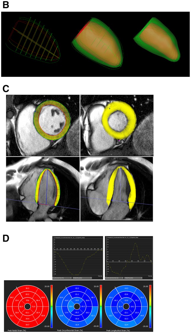
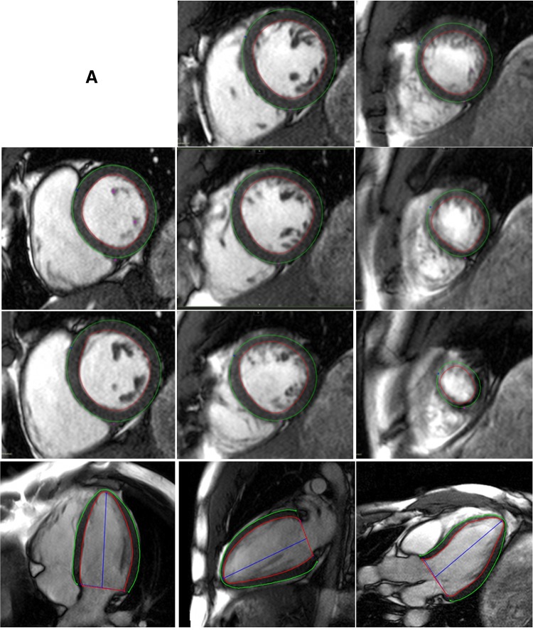
Fig. 1.

Steps taken for 3D FT-CMR. a Define endocardial and epicardial borders. b 3D construct of endocardial and epicardial borders are used to generate a 3D model of the myocardium in diastole which is tracked through to systole. c Ensure good quality tracking. d Results for global and/or segmental strain and strain rates

