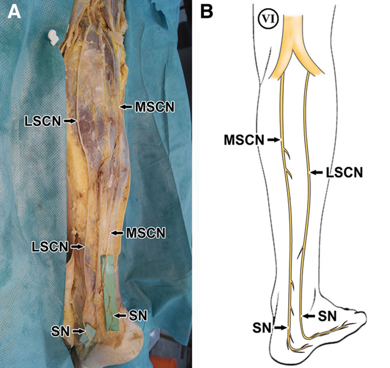
Fig. 1.

A, The LSCN and MSCN coursed in parallel, passed the retromalleolar region, and formed the SN separately in left leg. B, The new SN pattern drawing in the right leg, which was consistent with “Figure. 2” of the literature in the study by Riedl et al.
