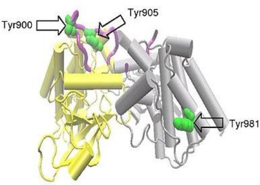
Fig. 1.

A RET dimer formed between two protein molecules, each spanning amino acids 703-1012 of the RET molecule and covering RET intracellular tyrosine kinase domain. One protein molecule, molecule A, is shown in yellow and the other, molecule B, in grey. The activation loop is colored purple and selected tyrosine residues in green. Part of the activation loop from molecule B is absent. mRET proto-oncogene with three main domains: an N-terminal extracellular domain with four cadherin-like repeats and a cysteine-rich region, a hydrophobic transmembrane domain, and a cytoplasmic tyrosine kinase domain. Phosphorylation of Tyr981 and the additional tyrosinesTyr1015, Tyr1062, and Tyr1096 are not covered by the above structure, though these have been shown to be important for initiation of the intracellular signal transduction processes[36].
