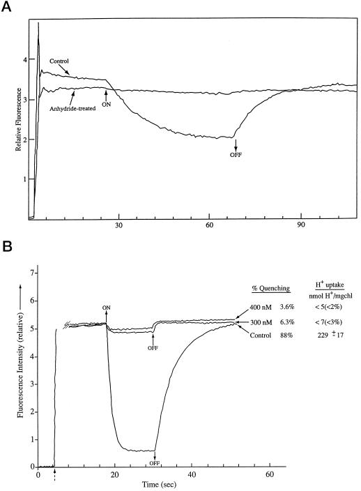
Figure 2.
A, ACMA fluorescence quenching in acetic-anhydride-treated and glycylglycine-quenched control spinach thylakoids. ACMA fluorescence quenching was measured under basal conditions (no ADP) as described in “Materials and Methods.” All measurements were done at 18°C. Light intensity was 40 and 180 μmol (m2 s)−1 in the control and anhydride-treated cases, respectively. The vertical line at approximately 2 s indicates the addition of the dye. B, ACMA fluorescence quenching in glycylglycine-quenched control spinach thylakoids with and without nigericin added compared with concurrent measurement of H+ uptake in parallel assays. ACMA procedures were as in Figure 1A. The dashed arrow indicates addition of the dye. For the H+ pump extent measurements, the assay conditions were the same as for the ACMA fluorescence assays except that 1 mm Tricine-KOH replaced the 50 mm Tricine-KOH to allow detection of pH changes, and 50 mm KCl was added to the pH assay mix to compensate for the lower Tricine-KOH. The photosynthetically active radiation (PAR) light intensity was measured at the rear of the cuvette compartment in the fluorimeter at full red light, and this intensity (40 μmol m−2 s−1) was reproduced at the front surface of the pH detection cuvette also using a red filter. The cuvette temperature was 18°C for both instruments. Glycylglycine (20 μg Chl mL−1)-quenched control thylakoids was used for both measurements. Duplicate measurements were made at the 300 and 400 nm nigericin levels (the same nigericin sample was used for both assays), and triplicate measurements were made for the control.

