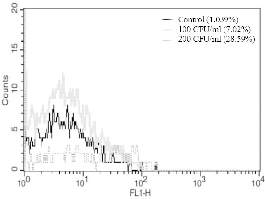
Fig. 6.

The fluorescent affinity of aptamer K3 to sterile CSF as the negative control (black curve), 200 CFU of N. meningitidis inoculated in CSF/ml (dark gray dotted), and 100 CFU of N. meningitidis inoculated in CSF/ml (light gray curve).

The fluorescent affinity of aptamer K3 to sterile CSF as the negative control (black curve), 200 CFU of N. meningitidis inoculated in CSF/ml (dark gray dotted), and 100 CFU of N. meningitidis inoculated in CSF/ml (light gray curve).