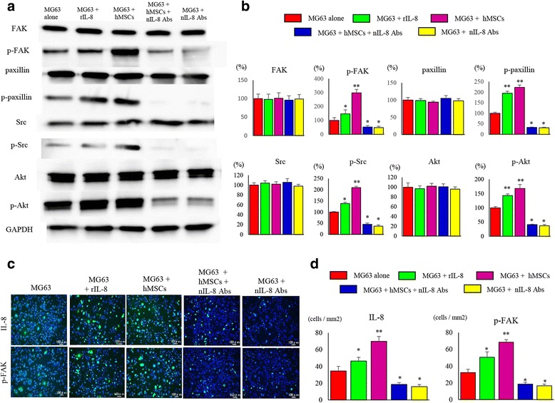
Fig. 6.

Changes in expression of FAK and its downstream factors related to invasive potential. a Changes in phosphorylation and the expression of protein factors relating to invasive potential were analyzed. Decreased phosphorylation of FAK, paxillin, Src, and Akt in MG63 cells was noted in the group administered nIL-8 Ab. b The quantification of western blot analysis. Data represents represent the mean ± SD of three independent experiments. p < 0.05 was considered to indicate significance: (*) p < 0.05, (**) p < 0.01. c Immunofluorescence staining of cultured MG63 cells showed decreased phosphorylation of FAK and paxillin in the group administered nIL-8 Ab. d The number of IL-8 and p-FAK positive cells per unit area. Data represents represent the mean ± SD of three independent experiments. p < 0.05 was considered to indicate significance: (*) p < 0.05, (**) p < 0.01
